Segmental arterial mediolysis (SAM) is a rare but important cause of abdominal haemorrhage and is usually treated with endovascular management. Surgical or conservative management can be performed depending on the patient’s situation. Since the course of conservative treatment is not well described in the existing literature, it must be used with caution. We describe the case of a 52-year-old man who was transferred to the emergency department because of abdominal pain, diarrhoea, and haematemesis. On arrival, he was haemodynamically unstable, with a blood pressure of 70/40 mmHg. After he was transfused and stabilised in the emergency department, contrast-enhanced computed tomography (CECT) revealed a haematoma around the transverse colon and ascites, without any evident extravasation. The peripheral branch of the middle colic artery (MCA) showed irregular calibre, suggesting SAM. Since the patient remained stable, we initially chose conservative management. However, a CECT scan performed on the third day of hospitalisation showed coexisting pulmonary thromboembolism (PTE). Because of the need for anticoagulation therapy, we performed open surgery. The pathological examination was consistent with SAM. Anticoagulation therapy was initiated the next day. A CECT scan performed on a postoperative day (POD) 13 showed no PTE. Although the patient defecated haemorrhagic stool and experienced hematemesis on PODs 19 and 23, respectively, colonoscopy, esophagogastroduodenoscopy, and repeated CECT scans revealed no evidence of rebleeding, and no recurrence was observed. Open surgery produced a relatively good postoperative course for the patient. Endovascular management, however, remains a reasonable first approach, considering its reportedly excellent outcomes.
Hinweise
Publisher’s Note
Springer Nature remains neutral with regard to jurisdictional claims in published maps and institutional affiliations.
Case Report
A 52-year-old man was transferred to the emergency department because of abdominal pain, diarrhoea, and haematemesis. He was haemodynamically unstable, with a blood pressure of 70/40 mmHg. The patient received fluid resuscitation and two units of red blood cells (RBCs), to stabilise him. Contrast-enhanced computed tomography (CECT) revealed a haematoma around the transverse colon and ascites without any evident extravasation (Fig. 1a). The peripheral branch of the MCA showed an irregular calibre (Fig. 1b), suggesting SAM. Since the patient remained stable, we chose conservative management.
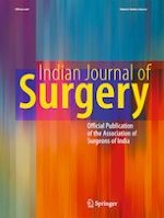
Fig. 1
CECT findings reveal a a haematoma inside the transverse mesocolon (yellow circle), ascites (yellow arrowheads), and b an irregular calibre of the peripheral branch of the middle colic artery with stenoses and dilations (red arrowheads). Another CECT reveals the PTE in the peripheral branch of the right lower pulmonary artery (c, blue arrowhead). The transverse mesocolon ruptured due to increased internal pressure (d, blue circle)
×
Three days later, he complained of chest pain. CECT showed a PTE in the peripheral branch of the right lower pulmonary artery (Fig. 1c). No exacerbation of the haematoma and ascites were observed. There was no deep vein thrombosis. Echocardiography showed no evidence of a cardiac cause of PTE; thus, we assumed transient hypercoagulation induced by haemorrhage could be responsible. Anticoagulation therapy was recommended; however, this could have led to an exacerbation of the intra-abdominal haemorrhage. Therefore, we decided to surgically remove the affected arteries.
Anzeige
When the abdominal cavity was opened, fresh blood was found throughout the upper abdomen. There was a haematoma inside the transverse mesocolon with the serosa ruptured because of increased internal pressure (Fig. 1d). We confirmed that the bleeding artery was completely torn. We resected the affected transverse colon and mesocolon to avoid rebleeding and end-organ ischaemia (Fig. 2a). The amount of bleeding was 1350 ml. Four units of RBCs were transfused during surgery. Pathological examination revealed dissection of the tunica media and intima, inside which a haematoma was present (Fig. 2b), establishing the final diagnosis of SAM.
Fig. 2
Resected specimen with a haematoma inside the mesocolon (a). The affected artery was torn apart, and arterial stumps were ligated (b, yellow allows). Pathological examination of the affected artery by haematoxylin and eosin staining reveals granulated tissue and thrombosis disrupting the tunica media (b)
×
Anticoagulation therapy was initiated the next day. CECT on POD 13 showed no recurrence. The postoperative course was uneventful until he defecated haemorrhagic stool and experienced haematemesis on PODs 19 and 23, respectively. Colonoscopy and esophagogastroduodenoscopy revealed no evidence of active bleeding, and no recurrence was observed. Repeated CECT showed no recurrence of abdominal haemorrhage and PTE until the patient was transferred for rehabilitation on POD 40.
Discussion
SAM was first reported in 1976 by Slavin and Gonzales [1], but the definitive aetiology is still unknown. Vessel injury caused by a vasospastic event—such as hypoxia, hypertension, vasopressor administration, or recent anaesthesia—may be a cause, resulting in the lysis and degeneration of the media of the arterial wall [2]. Pathological features in previous studies have shown that granulated tissue disrupts the tunica media without inflammatory cell infiltration and results in stenosis, occlusion, dissection, or aneurysm. Multiple small aneurysms alternating with intact arterial segments produce a “string of beads” sign on CECT or angiography [3]. Aneurysm rupture can be life-threatening, with an estimated mortality rate approaching 50% [4].
SAM usually involves the splanchnic arteries, such as the superior mesenteric artery, celiac axis, inferior mesenteric artery, and renal arteries. Sudden abdominal pain and haemodynamic shock are clues [5].
Anzeige
Fibromuscular dysplasia, connective tissue disorders, and infections are the differential diagnoses. Although some of these are challenging mimics, establishing the correct clinical diagnosis of SAM is not difficult with appropriate scrutiny of medical history, physical examinations, and investigation modalities [3, 5, 6].
Endovascular management usually provides better outcomes than surgery [3, 7], and conservative management is acceptable for haemodynamically stable patients [5, 6, 8]. However, short-interval follow-ups are essential when choosing conservative management.
We initially chose conservative management since the patient remained stable following resuscitation. However, the coexistence of SAM and PTE was challenging since it required simultaneous haemostasis and anticoagulation. There is currently no evidence for continuing conservative management when the risk of bleeding increases. Endovascular management was considered because of its excellent outcomes and minimal invasiveness. However, repeated CECT scans did not show any extravasation, and we anticipated difficulties in confirming the affected artery with angiography. Moreover, post-procedural anticoagulation therapy was immediately needed. Open surgery, while more invasive, provided the prompt and certain elimination of the affected area. While endovascular management is appropriate to consider, it should only be attempted with trained surgeons available for the surgery.
Retrospectively, the strategy for treating haemorrhage caused by SAM is simple: the first choice is endovascular management; should endovascular management be unavailable or fail, open surgery may be chosen, and conservative management may be a viable choice if the patient is hemodynamically stable and presents limited symptoms [7]. The first two approaches should be tried with surgical backup available in case of failure or rapid exacerbation.
Acknowledgements
We would like to thank Editage for English proofreading.
Declarations
Ethics Approval and Consent to Participate
This study was approved by the Ethics committee of Kurashiki Central Hospital.
Consent for Publication
Written informed consent was obtained from the patient for publication of this case report and any accompanying images.
Conflict of Interests
The authors declare no competing interests.
Anzeige
Open AccessThis article is licensed under a Creative Commons Attribution 4.0 International License, which permits use, sharing, adaptation, distribution and reproduction in any medium or format, as long as you give appropriate credit to the original author(s) and the source, provide a link to the Creative Commons licence, and indicate if changes were made. The images or other third party material in this article are included in the article’s Creative Commons licence, unless indicated otherwise in a credit line to the material. If material is not included in the article’s Creative Commons licence and your intended use is not permitted by statutory regulation or exceeds the permitted use, you will need to obtain permission directly from the copyright holder. To view a copy of this licence, visit http://creativecommons.org/licenses/by/4.0/.
Publisher’s Note
Springer Nature remains neutral with regard to jurisdictional claims in published maps and institutional affiliations.
Mit der Zeitschrift Die Chirurgie erhalten Sie zusätzlich Online-Zugriff auf weitere 43 chirurgische Fachzeitschriften, CME-Fortbildungen, Webinare, Vorbereitungskursen zur Facharztprüfung und die digitale Enzyklopädie e.Medpedia.
In der Notaufnahme wird die Chance, Opfer von häuslicher Gewalt zu identifizieren, von Orthopäden und Orthopädinnen offenbar zu wenig genutzt. Darauf deuten die Ergebnisse einer Fragebogenstudie an der Sahlgrenska-Universität in Schweden hin.
Darüber reden und aus Fehlern lernen, sollte das Motto in der Medizin lauten. Und zwar nicht nur im Sinne der Patientensicherheit. Eine negative Fehlerkultur kann auch die Behandelnden ernsthaft krank machen, warnt Prof. Dr. Reinhard Strametz. Ein Plädoyer und ein Leitfaden für den offenen Umgang mit kritischen Ereignissen in Medizin und Pflege.
Ein Frauenanteil von mindestens einem Drittel im ärztlichen Op.-Team war in einer großen retrospektiven Studie aus Kanada mit einer signifikanten Reduktion der postoperativen Morbidität assoziiert.
Bei schwerer Aortenstenose und obstruktiver KHK empfehlen die Leitlinien derzeit eine chirurgische Kombi-Behandlung aus Klappenersatz plus Bypass-OP. Diese Empfehlung wird allerdings jetzt durch eine aktuelle Studie infrage gestellt – mit überraschender Deutlichkeit.
Update Chirurgie
Bestellen Sie unseren Fach-Newsletterund bleiben Sie gut informiert.
Das Karpaltunnelsyndrom ist die häufigste Kompressionsneuropathie peripherer Nerven. Obwohl die Anamnese mit dem nächtlichen Einschlafen der Hand (Brachialgia parästhetica nocturna) sehr typisch ist, ist eine klinisch-neurologische Untersuchung und Elektroneurografie in manchen Fällen auch eine Neurosonografie erforderlich. Im Anfangsstadium sind konservative Maßnahmen (Handgelenksschiene, Ergotherapie) empfehlenswert. Bei nicht Ansprechen der konservativen Therapie oder Auftreten von neurologischen Ausfällen ist eine Dekompression des N. medianus am Karpaltunnel indiziert.
Das Webinar beschäftigt sich mit Fragen und Antworten zu Diagnostik und Klassifikation sowie Möglichkeiten des Ausschlusses von Zusatzverletzungen. Die Referenten erläutern, welche Frakturen konservativ behandelt werden können und wie. Das Webinar beantwortet die Frage nach aktuellen operativen Therapiekonzepten: Welcher Zugang, welches Osteosynthesematerial? Auf was muss bei der Nachbehandlung der distalen Radiusfraktur geachtet werden?
Inhalte des Webinars zur S1-Leitlinie „Empfehlungen zur Therapie der akuten Appendizitis bei Erwachsenen“ sind die Darstellung des Projektes und des Erstellungswegs zur S1-Leitlinie, die Erläuterung der klinischen Relevanz der Klassifikation EAES 2015, die wissenschaftliche Begründung der wichtigsten Empfehlungen und die Darstellung stadiengerechter Therapieoptionen.