Long-term clinical outcomes of Er:YAG or Er,Cr:YSGG lasers utilized as monotherapy or as adjuncts to mechanical therapy in the treatment of chronic periodontitis: a systematic review
verfasst von:
Triantafyllia Vagdouti, Charis Theodoridis, Georgia Tseleki, Ioannis Vouros
The aim of the present systematic review was to address the following focused question: In patients with generalized chronic periodontitis, what is the long-term effect of the Er:YAG or Er,Cr:YSGG lasers, as monotherapy or as adjuvant to mechanical therapy, on the following clinical outcomes: probing pocket depth (PPD), clinical attachment level (CAL), bleeding on probing (BOP), and gingival index (GI).
Methods
A thorough electronic search was performed in PubMed, Scopus, Cochrane, Web of Science, and Ovid databases according to PRISMA guidelines. The screening process and data extraction was conducted independently by two reviewers. A quality assessment using Cochrane Collaboration Methodology for randomized controlled trials (RCTs) was performed.
Results
Eight eligible RCTs fulfilled the criteria. Two RCTs utilising Er,Cr:YSGG laser, and six RCTs using Er:YAG laser in conjunction with non-surgical periodontal therapy. The primary outcome was PPD, while the secondary outcomes were CAL, BOP, and GI. When evaluating CAL benefits, two out of two of the included studies, which assessed Er:YAG as monotherapy in 24 months, indicated a significant difference in favor of Erbium lasers compared to SRP. It seems that Erbium lasers perform better in terms of PPD reduction compared to SRP in both 12-month and 24-month follow-up periods, especially with regard to moderate and deep periodontal pockets. The quality assessment revealed that four studies were presented with some concerns, while the rest of the studies were judged to be at low risk of bias.
Conclusion
It may be advocated that Er:YAG and Er,Cr:YSGG lasers as monotherapy or as adjunct to SRP seem to perform better in terms of CAL and PPD reduction in the long term, especially in deep pockets ≥ 7 mm; nevertheless, limited evidence for appropriate comparability is available in the existing literature.
Hinweise
The original online version of this article was revised due to errors in references.
Springer Nature remains neutral with regard to jurisdictional claims in published maps and institutional affiliations.
Introduction
Periodontitis is a chronic multifactorial bacterial–induced inflammatory disease characterized by progressive destruction of the tooth-supporting apparatus. Assessment of loss of supporting tissues is based on clinical attachment loss (CAL), probing pocket depth (PPD), bleeding on probing (BOP), and radiographically assessed alveolar bone loss [1]. Periodontal disease is a condition that undoubtfully affects patients’ well-being and its association with poor oral health-related quality of life (OHRQoL) has been well established, especially in moderate and severe disease forms [2, 3]. Furthermore, in a large-scale study, it has been shown that disease severity, in terms of higher staging and grading, is connected to a greater risk for tooth loss [4].
Cause-related periodontal therapy consists of subgingival instrumentation with or without adjunctive use of chemical agents, local or systemic antibiotics, and lasers. Different treatment modalities have been proposed aiming at biofilm control and the resolution of periodontal inflammation by using mechanical instrumentation, hand instruments, and ultrasonic devices. A short- and long-term beneficial effect of mechanical non-surgical therapy has been established by a significant number of clinical and microbiological studies [5, 6]. Suvan and coworkers have published a systematic review and meta-analysis, which included four RCTs, and found that there were no differences between ultrasonic and hand instruments, with both being equally effective. In general, 6 to 8 months following cause-related therapy, a mean reduction of PD of 1.5 mm was found at shallow (4–6 mm) probing pocket depths, while at deep sites (≥ 7 mm), a mean reduction of 2.6 mm was found [7]. Adjunctive products have been developed in order to improve the clinical outcome and avoid surgical intervention.
Anzeige
LASER is an acronym for Light Amplification by the Stimulated Emission of Radiation. Excited atoms from an active medium of a laser device emit photons, with the aid of a light source, in an intensifying manner and produce a single wavelength radiation [8]. The most commonly used lasers in periodontology are diode lasers (450–1064 nm), solid-state lasers, which include Neodymium YAG (Nd:YAG, 1064 nm), Erbium, Chromium YSGG (Er,Cr:YSGG, 2780 nm), Erbium YAG (Er:YAG 2940 nm), and laser gas CO2 (10,600 nm) [9].
Lasers were introduced in non-surgical periodontal therapy as adjunct approaches, in order to overcome difficulties in accessing deep pockets and furcation defects, provide better visualization through hemostasis, bactericidal effect, improve healing, and reduce the need for surgical intervention [10]. Moreover, a reduction of inflammatory mediators such as interleukin-1β, interleukin-6, tumor necrosis factor-α, matrix metalloproteinase-8, and reduction of the bacterial load has been shown [11‐13].
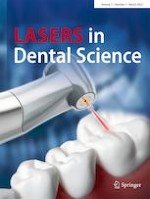
The Er:YAG and Er,Cr:YSGG lasers are hard tissue lasers, which interact with water and hydroxyapatite. Once these lasers mediate with water, the water evaporates and causes “microexplosions” leading to a calculus removal. Erbium lasers can ablate calculus efficiently by removing the smear layer and enhancing growth and adhesion of cells [14]. Moreover, it has been demonstrated in vitro that the Er:YAG laser removes subgingival calculus similarly to manual scaling and root planing, as well as, to ultrasonic scaling [15, 16]. The Er:YAG and Er,Cr:YSGG lasers can be also used for soft tissue debridement of the diseased epithelial lining [17] (Fig. 1), and they are considered as the most suitable lasers for periodontal therapy producing minimal thermal-related side effects [10]. Also, it has been shown in vitro cultures that erbium lasers are bactericidal against P. gingivalis and A. actinomycetemcomitans and they are effective in removing root endotoxins [18]. However, most of the clinical studies, although they have not made a thorough research on this, have not shown any significant difference between test and control groups with regard to bacterial load. Malali et al. showed a significant reduction at the levels of spirochetes and motile rods [19]. Numerous studies have shown a decrease in inflammation and probing pocket depth. Erbium lasers have been used in RCTs as monotherapy or as adjuncts to mechanical therapy. The studies up to now have not reached any concrete conclusion as to whether laser monotherapy is superior or, at least, comparable to the traditional periodontal therapy [20, 21]. Lin et al. performed a systematic review and meta-analysis on laser monotherapy, demonstrating that the use of the Er:YAG laser has similar results with the conventional mechanical therapy [22].
Fig. 1
Er,Cr:YSGG in non-surgical periodontal therapy
×
Until recently, four systematic reviews and meta-analyses have been conducted, focusing mostly on short-term results (≤ 3 months) [23‐26]. One systematic review and meta-analysis that refers to the Er,Cr:YSGG laser concluded that it might provide additional effectiveness in PD reduction and CAL gain, although statistically significant differences were found only in short-term level, as they did not include any long-term studies (≥ 6 months), and two meta-analyses have been conducted concerning Er:YAG lasers, stating that there were significant short-term differences, but not at medium or long-term evaluations [23, 25, 26]. Nevertheless, only two of the included clinical studies presented clinical outcomes in a long-term time frame. The consensus report from the American Academy of Periodontology, suggested that there is a modest additional clinical benefit from the adjunctive use of erbium laser, being more evident at deep periodontal pockets (≥ 7 mm) [20, 21]. In the same vein, Lavu and colleagues, who published an umbrella review and gathered all the information from the available systematic reviews, concluded that there was a lack of clinical data at > 6-month follow-up, but in the short-term, it appears that there are clinical benefits [27].
Anzeige
In the absence of scientific evidence on the use of erbium lasers in non-surgical periodontal therapy in the long-term, the present study was conducted with the purpose to present a systematic appraisal of the available literature using an evidence-based approach, which enrolls the 12-month and 24-month clinical outcomes of the Er:YAG or Er,Cr:YSGG laser as monotherapy or as adjunct to mechanical therapy in patients with chronic periodontitis.
Materials and methods
Design of the study
This systematic review was conducted according to the recommendations and principles of the Cochrane Collaboration [28], as well as the PRISMA statement [29]. Prior to study initiation, a comprehensive protocol was developed and approved by all authors in a commensurable methodological perspective as applied in previous works of our research team [30, 31]. This detailed protocol included several sections and research methods, including the search strategy, definition of eligibility, inclusion criteria, screening techniques, data extraction, quality assessment, and data presentation and assessment.
Focused question
In patients with untreated generalized chronic periodontitis, what is the long-term effect of Er:YAG or Er,Cr:YSGG lasers, as monotherapy or as adjunct to mechanical therapy, on the following clinical outcomes: probing pocket depth, clinical attachment level, bleeding on probing, and gingival index?
Inclusion criteria
1.
Randomized clinical trials (RCTs), controlled clinical trials (CCTs), cohort studies, and original prospective clinical studies, comparing the Er:YAG or Er,Cr:YSGG lasers with ultrasonic scaler and/or hand instruments and reporting numerical data on probing pocket depth, clinical attachment level, bleeding on probing, and gingival index.
2.
Single-arms of prospective clinical studies utilizing the Er:YAG or Er,Cr:YSGG lasers in patients with untreated periodontitis and reporting on the aforementioned clinical parameters.
3.
Both studies with split-mouth design and parallel design were considered for inclusion.
4.
All included studies have to report on a minimum follow-up period of 12 months.
5.
Included studies or arms of studies had to incorporate at least 15 periodontitis patients.
6.
Smokers were included.
Exclusion criteria
1.
Case reports, case series, reviews, editorials, retrospective studies, letters to the editor, personal opinions, book chapters, in vitro studies, short communications, conference abstracts, and animal studies were excluded.
2.
Studies or arms of studies presenting only SRP clinical outcomes, without the utilization of the Er:YAG, or the Er,Cr:YSGG were excluded.
3.
Studies in patients with medical conditions possibly affecting periodontal therapy such as cancer, uncontrolled diabetes mellitus, diseases affecting bone metabolism, and/or particular medication intake.
4.
Studies utilizing the Diode, CO2, Nd: YAG, KTP, Argon and other laser types, other than Er:YAG, or Er,Cr:YSGG or performing low-level laser therapy or photodynamic therapy.
5.
Studies reporting on < 12-month follow-up period were excluded as well.
Intervention types
All Er:YAG laser types and parameters, as well as the Er,Cr:YSGG laser types and parameters, having been utilized for periodontal therapy application were considered. With regard to scaling and root planing (SRP), either ultrasonic scalers or hand curettes, as well as combination of these, were regarded as SRP treatment modalities.
Compared population characteristics
Four population groups were created, with participants being adult patients with untreated generalized chronic periodontitis. The three out of the four groups were test groups: periodontitis patients having received Er:YAG radiation as monotherapy (T1), periodontitis patients having received the Er,Cr:YSGG laser as an adjunct to SRP (T2), and periodontitis patients been treated with the Er:YAG as an adjunct to SRP (T3). The fourth patient group comprised periodontitis patients, who were treated with SRP only and this group has been considered the control group.
Compared outcome measurements
Clinical probing pocket depth (PPD) was considered as the primary outcome variable in the present systematic review. PPD ought to have been measured and provided with numerical data by the included studies. Additional clinical parameters, such as clinical attachment level, bleeding on probing and gingival index were also obtained and considered as secondary outcomes.
Search strategy
The search strategy includes electronic databases, supplement by hand searches. A comprehensive electronic search was conducted by two authors (T.V. and G.T.) independently in MEDLINE via PubMed, Scopus, Cochrane Central Register of Controlled Trials (CENTRAL), Web of Science, and Ovid databases up to and including March 30th, 2022. A combination of MeSH terms and text words was utilized and the electronic search was formulated as follows:
Anzeige
Population/outcomes
Periodontitis or chronic periodontitis or periodontal inflammation or periodontal disease or periodontal therapy or periodontal treatment or non-surgical therapy or non-surgical treatment or scaling and root planing or SRP or dental scaling or root planing or periodontal debridement or periodontal pocket or probing pocket or clinical attachment level or attachment loss or bleeding on probing or periodontal parameters or alveolar bone loss.
Interventions
Lasers or solid-state lasers or solid state lasers or Er: yag or erbium yag or erbium yag laser or erbium-doped yttrium aluminium garnet laser or erbium lasers or Er,Cr: YSGG or erbium chromium yttrium scandium gallium laser or laser therapy or laser debridement or laser treatment or dental lasers.
Type of studies
In order to decrease the loss of any relevant studies, any search filter based on methodological concepts and on specific types of studies was avoided. For the same reason, no language restrictions were considered.
These terms were then combined as follows: population/exposure/outcomes and interventions.
Screening of the references of the included studies and other pertinent reviews on the topic was conducted as well. Additionally, the following journals were considered potentially significant and were hand-searched: the International Journal of Periodontics and Restorative Dentistry, Journal of Clinical Periodontology, Journal of Periodontology, Periodontology 2000, Lasers in Medical Science, and Lasers in Surgery and Medicine.
Screening process and data extraction
Standardized screening forms were prepared and used to record data from the studies screened at different stages. Following that, evidence tables were designed containing the data from the included studies. Two reviewers (T.V. and G.T.) performed the screening independently and in duplicate. Firstly, all titles resulting from the searches were screened to eliminate irrelevant publications and study types. Afterwards, studies were excluded on an abstract level. The final stage of screening involved full-text reading using a predetermined data extraction form to record data and confirm the eligibility of each study based on inclusion and exclusion criteria. The following data were extracted from the selected studies: journal, year of publication, type of study, population characteristics, inclusion criteria, number of patients, smokers, follow-up period, type and parameters of laser used, type of intervention, clinical parameters considered (PD, CAL, BOP, GI), numerical data, and study design. The level of agreement between the reviewers was calculated utilizing kappa score. During each stage, any disagreement was resolved by discussion, and if necessary, a third reviewer was consulted (C.T.). If consensus on the inclusion of an article was not achieved, the article was included in the next stage of screening.
Anzeige
Quality assessment of selected studies
Two review authors (T.V. and G.T.) independently assessed the risk of bias of each selected study. The methodological quality was assessed using the Cochrane Collaboration Methodology for randomized controlled trials, the ROBINS-I Tool (RoB 2.0). The RoB 2.0 assessment includes five domains: randomization process, deviations from intended interventions, missing outcome data, outcome measurement, and selection of the reported result. The overall risk of bias is judged as low, high, and with some concerns [32]. The authors of the selected articles were contacted by email in order to obtain missing or unclear data.
Results
Study characteristics
A total of 10,165 publications were obtained from the initial search. After removal of duplicates, 7853 articles were considered eligible. Initially, 7692 titles were excluded. Following the abstract screening, 153 articles were excluded as they did not fulfill the inclusion criteria. Finally, a total of eight articles fulfilled the inclusion criteria and were included in this systematic review [33‐40]. The overall kappa score was found to be 0.90, which is characterized by a satisfying agreement. A PRISMA flow diagram is presented in Fig. 2 with all the details of the screening process.
Fig. 2
PRISMA flow chart
×
The main characteristics of the eight included articles are summarized in Table 1. All included studies were RCTs, including six split-mouth RCTs [33‐37, 40], and two parallel-arm RCTs, conducted by the same research team [38, 39]. Seven RCTs were conducted in university settings, while one study was performed in a private office setting [35]. Six studies evaluated the long-term results of laser-assisted periodontal therapy at 12 months [34, 36‐40], and two studies at 24 months [33, 35].
Table 1
Characteristics of the included studies
Author/year
Study design
Recruitment
Type of laser/laser parameters
Evaluation intervals
Smokers
Age
Gender (male/female)
Intervention
Population
Clinical parameters
Schwarz et al. 2003 (a)
Split-mouth RCT
University of the Saarland, Germany
Er: YAG
Energy level 160 mJ/pulse
Frequency 10 Hz
Fiber tip (0.5 × 1.65 or 0.5 × 1.1 mm)
12 months
24 months
Excluded
28–79 years old
6:14
Test: Er: YAG
Control: SRP
20 patients
110 teeth
660 sites with PPD > 4 mm
PPD, CAL, GI, PI, BOP, REC
Schwarz et al. 2003 (b)
Split-mouth RCT
University of the Saarland, Germany
Er: YAG
Energy level 160 mJ/pulse
Frequency 10 Hz
Fiber tip (0.5 × 1.65 or 0.5 × 1.1 mm)
3 months
6 months
12 months
Unclear
28–79 years old
Test 1: Er: YAG
Test 2: Er: YAG + SRP
20 patients
100 teeth
600 sites with PPD > 4 mm
PPD, CAL, GI, PI, BOP, REC
Crespi et al. 2007
Split-mouth RCT
Private office, Italy
Er: YAG
energy level 160 mJ/pulse
Frequency 10 Hz
Energy density 94 J/cm2/pulse
Chisel quarz tip 400 μm
3 months
12 months
24 months
Unclear
37–65 years old
10:15
Test: Er: YAG
Control: ultrasonic scaler
25 patients
200 teeth
1200 sites with PPD > 4 mm
PPD, CAL, GI, PI
Lopes et al. 2010
Split-mouth RCT
Araraquara Dental School, Brazil
Er: YAG
energy level 100 mJ/pulse
Frequency 10 Hz
Energy density 12.9 J/cm2/pulse
Exposure duration
250–500 μs
Special application
Tip (1.1 × 0.5 mm)
1 month
3 months
6 months
12 months
Excluded
31–55 years old
7:14
Test 1: Er: YAG
Test 2: Er: YAG + SRP
Control 1: SRP
Control 2: No treatment
19 patients
76 teeth
76 sites with PPD 5–9 mm
PPD, CAL, GI, PI, BOP
Kelbauskiene et al. 2011
Split-mouth RCT
Kaunas University of Medicine, Lithuania
Er, Cr: YSGG
Average power 1W
Frequency 20 Hz;
pulse duration 140–200 μs;
fiber optic tip 600 μm
2 months
3 months
6 months
12 months
Excluded
26–58 years old
16:14
Test: Er, Cr: YSGG + SRP
Control: SRP
30 patients
Test: 135 teeth
509 sites
Control: 143 teeth
579 sites with PPD 3–6 mm
PPD, CAL, PI, BOP
Sanz- Sánchez et al. 2015
Parallel group
RCT
Universidad Complutense de Madrid, Spain
Er: YAG
energy level 160 mJ/pulse
Frequency 10 Hz;
sapphire tip
3 months
6 months
12 months
Included
25–80 years old
12:28
Test: Er: YAG + Ultrasonic scaler
Control: Ultrasonic scaler
Test: 19 patients (17 at 12 months)
Control: 21 patients (20 at 12 months)
25.4 mean number of teeth with PPD ≥ 4.5 mm
PPD, CAL, PI, BOP
Sanz-Sánchez et al. 2016
Parallel group
RCT
Universidad Complutense de Madrid, Spain
Er:YAG
energy level 160 mJ/pulse;
frequency 10 Hz;
sapphire tip (0.5 × 1.65 mm)
3 months
12 months
Included
25–80 years old
12:28
Test group: Er:YAG + Ultrasonic scaler
Control: Ultrasonic scaler
Test: 19 patients (17 at 12 months)
Control: 21 patients (20 at 12 months)
25.4 mean number of teeth with PPD ≥ 4.5 mm
PPD, PI, BOP, REC
Klokkevold et al. 2022
Split-mouth RCT
UCLA School of Dentistry, USA
Er,Cr:YSGG
Energy level 50 mJ/pulse
Average power 1.5W
Frequency 30 Hz
Radial Firing Perio Tip (RFTP5)
1 month
3 months
6 months
9 months
12 months
Excluded
27–65 years old
5:10
Test group: Er,Cr:YSGG + SRP
Control: SRP
15 patients
Test: 78 sites
Control: 77 sites with PPD ≥ 5mm
PPD, CAL, PI, BOP, REC
RCT randomized clinical trial, PPD probing pocket depth, CAL clinical attachment loss, GI gingival index, PI plaque index, BOP bleeding on probing, REC recession
Population characteristics
The sample size varied from 15 to 40 patients, 25 to 80 years old, and females were twice than males. All studies reported on patients diagnosed with generalized chronic periodontitis. Smokers were included in two articles [38, 39], excluded in four articles [33, 36, 37, 40], and was unclear whether they were included or not in two articles [34, 35]. Two studies included solely test group T1 and their respective control groups [33, 35]. Two studies included only test group T3 and one control group [38, 39], while two studies included only test group T2 and one control group [37, 40]. One study had two experimental groups T1 and T3, without any control group [34], and another study included two experimental groups T1 and T3 and one control group [36]. The control group in every study involved either an ultrasonic scaler or scaling and root planing.
Anzeige
Intervention characteristics
Er,Cr:YSGG laser-assisted periodontal therapy was performed in two studies [37, 40], whereas the Er:YAG laser-assisted periodontal therapy in six studies [33‐36, 38, 39]. Regarding laser parameters, five studies used an energy level of 160 mJ/pulse and a frequency of 10 Hz [33‐35, 38, 39], while the other studies ranged from 50 to 100 mJ/pulse and a frequency from 10 to 30 Hz [36, 37, 40]. On the other hand, diversity was found in fiber types and diameters.
Outcome characteristics
All included studies reported probing pocket depth (PPD) in mm. Clinical attachment loss, measured in mm, was provided in all studies except for one [38]. Bleeding on probing (BOP), calculated in percentages, was reported in all studies except for one [34]. Gingival index (GI) was presented in four articles [32‐35].
The clinical outcomes in terms of mean PPD values (± SDs) of the included studies are presented in Table 2. A statistically significant difference was reported in favor of the test groups in four studies, at 12 and 24 months [33, 35, 37, 39]. Two of the above studies reported a statistical significant difference in favor of the T1 group (Dif. 0.3–2.6 mm, p < 0.05), while the respective parameter was in favor of the T2 group (Dif 0.8 mm, p < 0.001) in another, and in favor of the T3 group (Dif 0.4 mm, p < 0.01) in the fourth trial [33, 35, 37, 39].
Table 2
Clinical outcomes of mean probing depths (PPD) and standard deviations (SD) of the included studies
Mean PPD (mm) SD
Dif (mm/%)
P value
Groups
T1: Er:YAG
T2: Er,Cr: YSGG + SRP test
T3: Er:YAG + SRP
Control (SRP)
Test
Control
Studies
Baseline
12 months
24 months
Baseline
12 months
24 months
Schwarz et al. 2003 (a)
T1
4.9 ± 0.7
3.0 ± 0.8
3.3 ± 0.9
5.0 ± 0.6
3.5 ± 1.3
3.7 ± 0.7
1.6/39
1.3/29.8
< 0.01
Schwarz et al. 2003 (b)
T1
5.0 ± 0.7
3.3 ± 0.7
-
-
-
-
1.7/40.9
-
NS
T3
5.2 ± 0.8
3.2 ± 0.8
-
2/47.6
-
Crespi et al. 2007
T1
PPD 5–6 mm: 5.49 ± 0.27
PPD ≥ 7mm: 7.92 ± 0.78
PPD 5–6 mm: 2.60 ± 0.37
PPD ≥ 7mm: 3.11 ± 0.41
PPD 5–6 mm: 2.61 ± 0.54
PPD ≥ 7mm: 3.05 ± 0.53
PPD 5–6 mm: 5.12 ± 0.39
PPD ≥ 7mm: 7.13 ± 0.53
PPD 5–6 mm: 4.02 ± 0.65
PPD ≥ 7mm: 4.820.37
PPD 5–6 mm: 4.120.74
PPD ≥ 7mm: 4.85 ± 0.64
PPD 5–6 mm: 2.88/71.1
PPD ≥ 7mm: 4.87/88.7
PPD 5–6 mm: 1.00/21.6
PPD ≥ 7mm: 2.28/38
PPD 5–6 mm: < 0.01
PPD ≥ 7mm: < 0.001
Lopes et al. 2010
T1
6.42 ± 1.1
4.76 ± 1.2
-
6.87 ± 1.1
4.58 ± 1.3
-
1.66/29.6
2.29/40
NS
T3
6.48 ± 1.2
4.29 ± 1.5
-
2.19/40.6
Kelbauskiene et al. 2011
T2
4.33 ± 1.08
2.75 ± 1.09
-
4.77 ± 0.79
3.31 ± 1.0
-
1.71/44.6
0.89/36.1
< 0.001
Sanz- Sánchez et al. 2015
T3
3.07 ± 0.31
2.48 ± 0.37
-
3.11 ± 0.32
2.71 ± 0.36
-
0.52/21.2
0.36/13.7
NS
Sanz-Sánchez et al. 2016
T3
6.01 ± 0.74
3.96 ± 0.78
-
6.02 ± 1.0
4.43 ± 1.02
-
1.97/41.1
1.50/30.4
0.01
Klokkevold et al. 2022
T2
6.1 ± 0.88
4.2 ± 0.98
-
6.2 ± 1.11
4.3 ± 1.23
-
1.9/36.8
1.9/36.1
0.487
NS not significant
Dif refers to the inter-group statistically significant differences from baseline to either 12 or 24 months
P value refers to statistically significant difference between groups
The clinical outcomes concerning the mean CAL values and their SDs of the included studies are depicted in Table 3. A statistically significant difference in favor of the T1 group was observed in two studies (Dif 0.7–3.02 mm, p < 0.05), while the respective CAL difference was in favor of the T2 group in another study (Dif 0.84 mm, p < 0.001), at 12 or 24 months, respectively [33, 35, 37]. The remaining five studies did not reveal any significant differences between test and control groups.
Table 3
Clinical outcomes of mean clinical attachment loss (CAL) and standard deviations (SD) of the included studies
Mean CAL (mm) SD
Dif (mm/%)
P value
Groups
T1: Er:YAG
T2: Er,Cr: YSGG + SRP test
T3: Er:YAG + SRP
Control (SRP)
Test
Control
Studies
Baseline
12 months
24 months
Baseline
12 months
24 months
Schwarz et al. 2003 (a)
T1
6.3 ± 1.1
4.5 ± 1.3
4.9 ± 1.0
6.5 ± 1.0
5.6 ± 1.4
5.8 ± 1.0
1.4/25
0.7/11.3
< 0.001
Schwarz et al. 2003 (b)
T1
5.0 ± 0.7
3.3 ± 0.7
-
-
-
-
1.7/40.9
-
NS
T3
6.6 ± 1.1
5.0 ± 0.7
-
1.6/27.5
-
Crespi et al. 2007
T1
PPD 5-6 mm: 6.27 ± 0.51
PPD ≥ 7mm: 8.41 ± 0.47
PPD 5-6 mm: 3.32 ± 0.64
PPD ≥ 7mm: 3.31 ± 1.01
PPD 5-6 mm: 3.35 ± 0.91
PPD ≥ 7mm: 3.11 ± 0.41
PPD 5-6 mm: 6.18 ± 0.42
PPD ≥ 7mm: 8.35 ± 0.33
PPD 5-6 mm: 4.89 ± 0.55
PPD ≥ 7mm: 6.33 ± 0.61
PPD 5-6 mm: 3.38 ± 0.79
PPD ≥ 7mm: 6.34 ± 0.92
PPD 5-6 mm: 2.92/60.7
PPD ≥ 7mm: 5.03/92
PPD 5-6 mm: 1.32/58.5
PPD ≥ 7mm: 2.01/27.3
PPD 5-6 mm: < 0.01
PPD ≥ 7mm: < 0.001
Lopes et al. 2010
T1
6.61 ± 1.1
5.93 ± 1.1
-
7.20 ± 1.3
5.79 ± 1.3
-
0.68/10.8
1.41/21.7
NS
T3
6.71 ± 1.4
5.56 ± 1.4
-
1.15/18.7
Kelbauskiene et al. 2011
T2
4.47 ± 1.2
2.8 ± 1.27
-
4.23 ± 0.92
3.4 ± 1.19
-
1.68/45.9
0.84/21.7
< 0.001
Sanz-Sánchez et al. 2015
T3
3.8 ± 0.74
3.44 ± 0.63
-
3.77 ± 0.46
3.57 ± 0.58
-
0.28/9.9
0.15/5.4
NS
Sanz-Sánchez et al. 2016
T3
-
-
-
-
-
-
-
-
-
Klokkevold et al. 2022
T2
6.8 ± 1.62
5.3 ± 1.57
-
6.9 ± 1.71
5.5 ± 1.76
-
1.5/24.7
1.4/22.5
0.767
NS not significant
Dif refers to the inter-group statistically significant differences from baseline to either 12 or 24 months
P value refers to statistically significant difference between groups
Mean BOP is presented in Table 4. Only two studies reported a significant difference among groups, one in favor of the T1 group (Dif 12%, p < 0.05), and one in favor of the T2 group (Dif 22%, p < 0.001) [33, 37]. Nonetheless, descriptive statistics of the GI presented in Table 5 did not show any significant difference between groups, although only four studies presented data for GI [33‐36].
Table 4
Clinical outcomes of mean bleeding on probing (BOP) and standard deviations (SD) of the included studies
Mean BOP in %
Dif (mm/%)
P value
Groups
T1: Er:YAG
T2: Er,Cr: YSGG + SRP test
T3: Er:YAG + SRP
Control (SRP)
Test
Control
Studies
Baseline
12 months
24 months
Baseline
12 months
24 months
Schwarz et al. 2003 (a)
T1
56
14
20
52
26
28
36
24
< 0.05
Schwarz et al. 2003 (b)
T1
61
16
-
-
-
-
45
-
NS
T3
58
14
-
44
-
Crespi et al. 2007
T1
-
-
-
-
-
-
-
-
-
Lopes et al. 2010
T1
100
39.7
-
100
32.3
-
60.3
67.7
NS
T3
100
33
-
67
Kelbauskiene et al. 2011
T2
79
9.8
-
74.1
26.7
-
69.2
47.4
< 0.001
Sanz- Sánchez et al. 2015
T3
64.41
28.57
-
65.44
30.61
-
31
35
NS
Sanz-Sánchez et al. 2016
T3
100
38
-
100
41
-
62
59
NS
Klokkevold et al. 2022
T2
78.21
55.84
-
71.43
63.64
-
22.37
7.79
0.198
NS not significant
Dif refers to the inter-group statistically significant differences from baseline to either 12 or 24 months
P value refers to statistically significant difference between groups
Table 5
Clinical outcomes of mean gingival index (GI) and standard deviations (SD) of the included studies
Mean GI SD
Dif (mm/%)
P value
Groups
T1: Er:YAG
T2: Er,Cr: YSGG + SRP test
T3: Er:YAG + SRP
Control (SRP)
Test
Control
Studies
Baseline
12 months
24 months
Baseline
12 months
24 months
Schwarz et al. 2003 (a)
T1
1.9 ± 0.6
0.4 ± 0.3
1.0 ± 0.6
1.9 ± 0.6
0.5 ± 0.3
1.1 ± 0.6
0.9/62
0.8/53.3
NS
Schwarz et al. 2003 (b)
T1
1.9 ± 0.7
0.6 ± 0.4
-
-
-
-
1.3/104
-
NS
T3
1.8 ± 0.6
0.5 ± 0.5
-
1.3/113
-
Crespi et al. 2007
T1
1.75 ± 0.58
0.64 ± 0.42
1.09 ± 0.61
1.75 ± 0.58
0.63 ± 0.35
1.01 ± 0.76
0.66/46.4
0.74/53.6
NS
Lopes et al. 2010*
T1
33.3
-
-
42.7
23.5
-
-
19.2
NS
T3
52.4
20.1
-
32.3/89.1
Kelbauskiene et al. 2011
T2
-
-
-
-
-
-
-
-
-
Sanz- Sánchez et al. 2015
T3
-
-
-
-
-
-
-
-
-
Sanz-Sánchez et al. 2016
T3
-
-
-
-
-
-
-
-
-
Klokkevold et al. 2022
T2
-
-
-
-
-
-
-
-
-
NS not significant
Dif refers to the inter-group statistically significant differences from baseline to either 12 or 24 months
P value refers to statistically significant difference between groups
*Lopes et al. [36] provided GI results in a presentative manner, in %
In four out of six studies reporting on the use of the Er:YAG laser [33, 35, 38, 39], and one out of two studies reporting on the use of the Er,Cr:YSGG as adjuncts in non-surgical therapy (T2 group), a statistically significant CAL gain and PPD reduction favoring the laser groups was found [37]. In this respect, the difference between the test and control groups may be limited, but it is noteworthy that it was considered significant at moderate and deep pockets (Table 6).
Table 6
Clinical outcomes in deep pockets (≥ 7 mm)
Authors
Groups
Clinical outcome
Follow-up time
P Value
Schwarz et al. 2003(a)
T1
1.4 mm greater CAL gain in favor of Er:YAG laser
24 months
p < 0.001
Crespi et al. 2007
T1
3.23 mm greater CAL gain and 1.8 mm more PPD reduction in favor of Er:YAG laser
12,24 months
p < 0.001
Klokkevold et al. 2022
T2
36.04% decrease in deep sites in favor of Er,Cr:YSGG laser + SRP
12 months
p = 0.044
Sanz-Sánchez et al. 2015
T3
5.62% decrease in deep sites in favor of the Er:YAG laser + SRP
12 months
p = 0.004
Sanz-Sánchez et al. 2016
T3
0.47 mm reduction in PPD in favor of the Er:YAG + SRP
12 months
p = 0.01
Quality assessment
The risk of bias of the included studies is presented in Fig. 3. Four of the included studies were considered to be with some concerns [33‐36], because of the randomization process and deviations from intended interventions. The rest of the studies were judged to be at low risk of bias [37‐40].
Fig. 3
Risk of bias in each domain and overall risk of bias, for all included studies, according to the Cochrane Collaboration Methodology for RCTs
×
Discussion
Laser-assisted periodontal therapy was introduced in non-surgical periodontal treatment as a method to achieve better outcomes than traditional therapy, improve healing, and reduce the need for future surgical interventions. Its use has been suggested as an alternative treatment modality in medically compromised patients who cannot undergo surgical periodontal treatment. Erbium family lasers have been shown to be efficacious for non-surgical periodontal treatment [11, 41]. Furthermore, cell adherence and re-attachment of periodontal ligament cells to the root surface, elimination of root endotoxins, and effective removal of calculus without causing melting or carbonization have been achieved [12, 15, 16, 42, 43].
The present systematic review focused on the long-term clinical evidence of Erbium lasers, as it appears that there is a lack of evidence, respectively. The aim of this systematic review was to assess the clinical outcomes of the Er:YAG and Er,Cr:YSGG laser as monotherapy or as adjuncts to SRP for chronic periodontitis treatment in the long-term (12 months and 24 months). The effectiveness of these lasers was evaluated through eight RCTs with a total of 209 patients. The clinical outcomes considered for evaluation were PPD, CAL gain, BOP, and GI.
Due to the fact that a great heterogeneity, with a non-comparable design, was observed among the studies, a meta-analysis could not be performed. Most of the included studies did not provide PPD changes and CAL changes, while the level of the analysis (patient/teeth/site) was different. In order to conduct a meta-analysis, the correlation coefficient r needs to be acknowledged. Therefore, we decided not to perform a value amputation and assume that r is 0.5, as this assumption would render the meta-analysis and its results questionable.
Findings from this systematic review indicate that there is a statistically significant difference in PPD in the long term in four studies (Dif. 0.3–3.07 mm, p < 0.05), [33, 35, 37, 39] in favor of the Erbium lasers, while a statistical significance in CAL gain was found in three studies (Dif 0.7–3.02 mm, p < 0.05) [33, 35, 37]. When evaluating CAL changes, two out of two of the included studies, which assessed Er:YAG as monotherapy in 24 months, indicated a significant difference in favor of Erbium lasers compared to SRP. It seems that Erbium lasers perform better in terms of PPD compared to SRP in both 12-month and 24-month follow-up periods, especially with regard to moderate and deep periodontal pockets.
Although the included studies in terms of laser parameters and smoking habits may be not homogenous and also taking into account that the number of the long-term studies are limited, the clinical outcomes of the present systematic review are encouraging, especially in moderate and deep pockets. Five of the studies included, analyzed the clinical outcomes in moderate and deep pockets, and showed that the Erbium lasers were performing better than SRP in deep pockets [33, 35, 38‐40] (Table 6). More specifically, two studies concerning Er:YAG laser as monotherapy found statistically significant differences in favor of the laser group. Schwarz et al. found a difference in CAL gain of 1.4 mm in 24 months, while Crespi et al. found a 3.23-mm difference at 24 months and a 1.8-mm difference in PPD (p < 0.001) for the laser group [33, 35]. As far as the adjunctive use of Er:YAG laser is concerned, Sanz-Sánchez and co-workers found that initially deep pockets in the laser group showed a 5.62% lower percentage of sites ≥ 4.5 mm at 12 months (p = 0.004) compared to the SRP group. In a similar trial, a 0.47-mm difference in PPDs in favor of the laser group for sites with deep pockets was observed at 12 months (p = 0.01) [38, 39]. Furthermore, in a more recent trial, a more significant pocket closure rate (PD ≤ 4 mm) of 36.04% of the sites with deep pockets ≥ 7 mm was observed at 12 months in the combined SRP/Er,Cr:YSGG Laser group compared to mechanical treatment only (p = 0.044) [40]. These results are in accordance with the consensus report from the American Academy of Periodontology, which states that the additional clinical benefit of Erbium lasers, as adjuncts to conventional therapy, is greater in deep pockets [20].
Another interesting issue in the implementation of Lasers would be the need for further surgical management of residual pockets after non-surgical treatment. Hence, achievement of residual pockets ≤ 5 mm presented with absence of bleeding would be advantageous thus avoiding the requirement for surgical approach. Accordingly, two of the included studies analyzing the residual PPDs found a significantly greater amount of PPDs > 5 mm in the control groups in comparison to the T3 group (73.08% vs 37.04%), as well as to the T2 group (17/27 sites vs 7/26 sites) at 12 months [38, 40].
Quality assessment of the included RCTs was performed using the ROBINS-I Tool. The assessment revealed that four out of eight studies presented with some concerns, mostly due to the randomization process and deviations from intended interventions, while the other four studies were considered at low risk of bias.
Hence, divergent results were found due to the high heterogeneity among studies. The included studies utilized different clinical parameters in order to evaluate the treatment outcome. More specifically, although all studies analyzed the mean PPD, one study performed a classification in shallow and deep pockets and analyzed the results separately without presenting the total PPD [35]. Further to mean PPD clinical outcomes, mean CAL outcomes were calculated in all studies but one [39]. Moreover, mean BOP percentage was not mentioned in one study [35] and mean GI measurements were mentioned in only four studies [37‐40].
It is noteworthy that there is a heterogeneity in baseline characteristics concerning the degree of periodontal disease severity in the included studies, a fact which leads to a different degree of PD or CAL reduction, rendering the results of those studies not comparable. Taken that into account it may be recommended, instead of assessing mean values of the clinical parameters to calculate the percentages of the values changes, thus allowing for a more objective comparison of treatment outcome among studies.
Another issue that should be addressed is the diversity in laser settings (power settings, energy level, frequency, pulse duration, time, proximity of the tip to the root surface, angulation of the tip) applied in the studies. A cause of concern in this respect may be lack of competence in laser basic principles and objectives, which is crucial for safely and successfully operating a laser device. A thorough and profound training in lasers biophysics is a conditio sine qua non before performing any laser therapy in humans.
All four systematic reviews and meta-analyses published so far are indicating that the added clinical benefit of the Erbium lasers is evidence based only in the short-term [23‐26]. One systematic review and meta-analysis concerning the use of Er,Cr:YSGG laser in non-surgical periodontal therapy did not include any studies in the long term [26]. As far as the Er:YAG laser is concerned, one systematic review and two meta-analyses included only two and three long-term studies respectively that resulted in insignificant statistical differences between groups in PPD reduction and CAL gain [23‐25]. The interpretation of the results of previous systematic reviews is based on the assumption that the effect of laser therapy diminishes with time, laser protocols are inconsistent and inclusion criteria particularly related to smoking habits are unclear. On the other hand, a small number of long-term studies was included, without taking into consideration all the available literature, in contrast to our systematic review, that included a thorough electronic research and was accomplished according to PRISMA guidelines. Moreover, as we mentioned above, the most evident clinical benefit that emerges from our systematic review is the improvement of the clinical parameters in deep pockets. This is an important parameter that is not evaluated in the previous systematic reviews. Additionally, the baseline PPDs in most of the included studies, comprised mainly shallow to moderate pockets, with a small percentage of deep pockets, a fact that might explain the lack of clinical significance of the findings.
However, there are some limitations in the present study, which should be mentioned. The most important limitation is that six out of eight studies were conducted as split-mouth. Split-mouth studies may distort the outcome as they can cause a wash-over effect and therefore should be avoided. Furthermore, there is a lack of standard laser settings (energy level, frequency, pulse duration, tip, frequency of application) and every researcher chooses to apply empirically a specific protocol for the reasons we have discussed above. Smoking is an evidenced based risk factor in periodontal disease affecting the final study outcomes. And of course, we cannot fail to mention the increased cost of the laser devices, although there is no cost-effectiveness analysis available to evaluate cost benefit rate [10, 44].
Conclusions
This systematic review provides limited evidence on the use of Er:YAG and Er,Cr:YSGG laser as monotherapy or as an adjunct to SRP in the long-term. Nevertheless, concerning PPD reduction and CAL gain, Erbium lasers seem to offer an improved clinical outcome in the long term. There is evidence that Erbium lasers are effective especially in deep pockets ≥ 7 mm, but due to increased heterogeneity and limitations in sample size among the original clinical studies, the results should be interpreted with caution.
Future research recommendations
As the evidence on the use of erbium lasers in periodontal therapy is still lacking, further research is needed. First of all, a parallel-arm randomized controlled clinical trials should be conducted, with a long-term follow-up, larger sample size, and standardized laser parameters and protocols. A more consistent methodology in data reporting and a consensus on the laser protocol should be implemented in order to reduce heterogeneity of the studies. Secondly, the clinical outcomes of PPDs and CAL gain should be presented as mean and change values, but also in subgroups of moderate and deep pockets, so that conclusions can be drawn on the prevention of surgical periodontal treatment. For the same reason, it would be advisable to report the residual PPDs, in order to evaluate the clinical effectiveness of the Er:YAG and Er,Cr:YSGG laser. Thus, more recommendations can be made on the basis of scientific evidence. Finally, a cost-effectiveness analysis would aid in the evaluation of the erbium lasers.
Declarations
Ethical approval
Not applicable.
Informed consent
Not applicable.
Conflict of interest
The authors declare no competing interests.
Open AccessThis article is licensed under a Creative Commons Attribution 4.0 International License, which permits use, sharing, adaptation, distribution and reproduction in any medium or format, as long as you give appropriate credit to the original author(s) and the source, provide a link to the Creative Commons licence, and indicate if changes were made. The images or other third party material in this article are included in the article's Creative Commons licence, unless indicated otherwise in a credit line to the material. If material is not included in the article's Creative Commons licence and your intended use is not permitted by statutory regulation or exceeds the permitted use, you will need to obtain permission directly from the copyright holder. To view a copy of this licence, visit http://creativecommons.org/licenses/by/4.0/.
Publisher's note
Springer Nature remains neutral with regard to jurisdictional claims in published maps and institutional affiliations.
Long-term clinical outcomes of Er:YAG or Er,Cr:YSGG lasers utilized as monotherapy or as adjuncts to mechanical therapy in the treatment of chronic periodontitis: a systematic review
verfasst von
Triantafyllia Vagdouti Charis Theodoridis Georgia Tseleki Ioannis Vouros
Sie sei „ethisch geboten“, meint Gesundheitsminister Karl Lauterbach: mehr Transparenz über die Qualität von Klinikbehandlungen. Um sie abzubilden, lässt er gegen den Widerstand vieler Länder einen virtuellen Klinik-Atlas freischalten.
Gesundheitsminister Lauterbach hat die vom Bundeskabinett beschlossene Klinikreform verteidigt. Kritik an den Plänen kommt vom Marburger Bund. Und in den Ländern wird über den Gang zum Vermittlungsausschuss spekuliert.
In einer Leseranfrage in der Zeitschrift Journal of the American Academy of Dermatology möchte ein anonymer Dermatologe bzw. eine anonyme Dermatologin wissen, ob er oder sie einen Patienten behandeln muss, der eine rassistische Tätowierung trägt.
Extreme Arbeitsverdichtung und kaum Supervision: Dr. Andrea Martini, Sprecherin des Bündnisses Junge Ärztinnen und Ärzte (BJÄ) über den Frust des ärztlichen Nachwuchses und die Vorteile des Rucksack-Modells.
Update Zahnmedizin
Bestellen Sie unseren kostenlosen Newsletterund bleiben Sie gut informiert – ganz bequem per eMail.